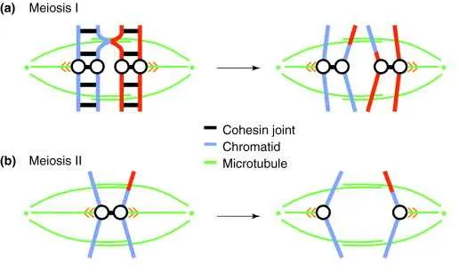
Look for the stage where homologous chromosomes, each consisting of two sister chromatids, are pulled toward opposite poles. This phase is marked by the movement of paired chromosomes separating, but the sister chromatids remain connected at their centromeres.
Key features include: visible spindle fibers attached to centromeres, clear separation of homologous pairs without splitting chromatids, and chromosomes migrating away from the metaphase plate.
Images depicting chromatids already divided or individual chromatids moving apart correspond to later stages. The correct visual will show chromosome pairs distinctly moving to poles, not single chromatids.
Identification of Stage I Chromosome Separation

The correct visual depiction of the first division’s chromosome segregation shows homologous pairs pulled apart toward opposite poles. Key features include chromosomes still composed of two sister chromatids, moving as distinct units, unlike the subsequent phase where chromatids separate individually.
Look for paired homologues aligned on the metaphase plate being pulled apart, with spindle fibers visibly attaching to centromeres. The image should exclude single chromatids separating, confirming the event belongs to the initial reductional division rather than the second division of the process.
Visual clues include elongated chromosome structures moving toward cell poles, with no chromatids splitting at centromeres. This confirms the stage when homologous chromosomes, not chromatids, are segregated, reducing chromosome number by half in each daughter cell.
Key Visual Features to Identify Anaphase I in Diagrams
Focus on the following characteristics to accurately distinguish the stage of the first division in meiosis:
- Homologous chromosome pairs move toward opposite poles; sister chromatids remain joined at their centromeres.
- Each pole displays a complete set of duplicated chromosomes, but they are still in their replicated form.
- The spindle fibers are visibly attached to centromeres, pulling the bivalents apart.
- No separation of sister chromatids occurs at this point, differentiating it from the later stage of division.
- Chromosomes appear thicker and shorter than during earlier phases, with clear movement away from the metaphase plate.
- Cell shape elongates as the spindle apparatus pushes poles apart, visible as stretched cytoplasm between chromosome clusters.
When examining images, confirm these traits together rather than relying on one single feature for correct identification.
Common Misinterpretations When Distinguishing Anaphase I Diagrams
Focus on the separation of homologous chromosome pairs rather than sister chromatids, as the former move to opposite poles during this stage. Confusing the splitting of sister chromatids with the segregation of homologues leads to incorrect identification.
Pay attention to the number of chromosomes at each pole; cells should show paired chromatids moving apart, not individual chromatids as in later phases. If the image depicts sister chromatids separating, it corresponds to a different division step.
Beware of illustrations that display chromosomes aligned on a single plane without visible tension or movement towards poles, as this suggests metaphase rather than the stage of homologous separation.
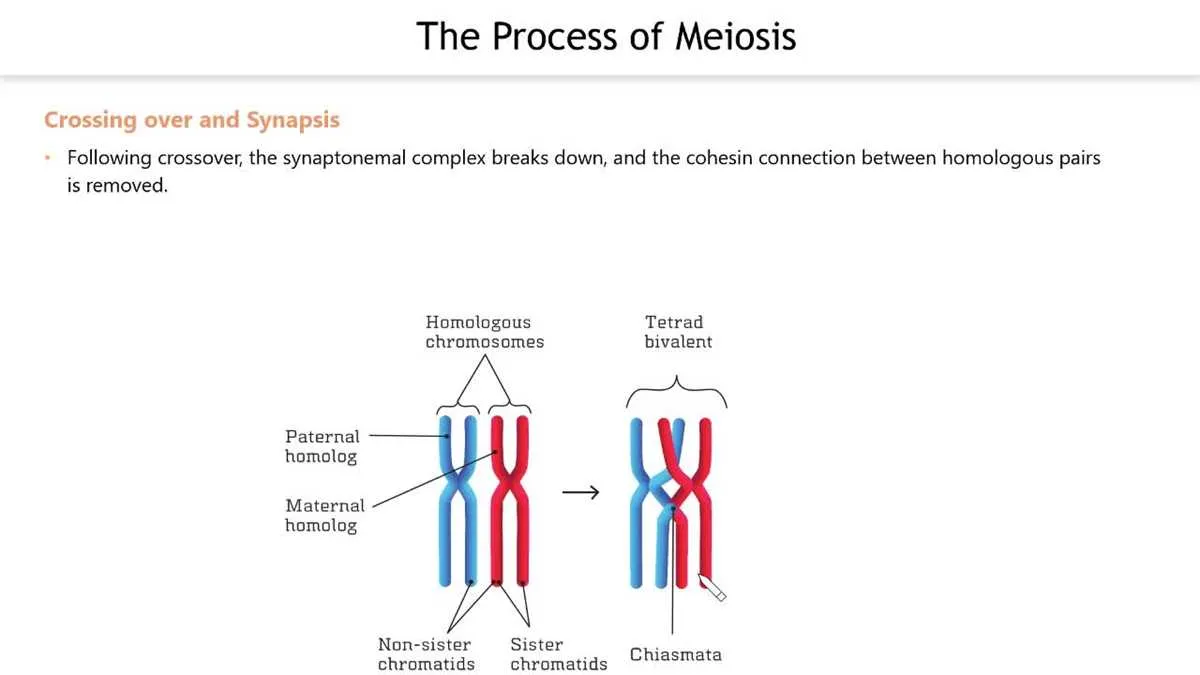
Check for the presence of chiasmata or crossover points still holding sister chromatids together; their disappearance indicates progression beyond the first division phase. Misreading these structures can cause confusion about the cell cycle stage.
Note spindle fiber attachment sites: during this phase, microtubules connect to homologous chromosome pairs, not individual chromatids. Mistaking these connections affects correct stage recognition.
Lastly, confirm that the cell remains diploid with duplicated chromosomes rather than haploid; the halving of chromosome number occurs only after this segregation, so any depiction suggesting haploidy is misleading for this division point.
Step-by-Step Guide to Analyzing Cellular Division Illustrations for Phase I Separation
Identify homologous chromosome pairs aligned at the cell’s equatorial plane. In this stage, each pair remains connected as tetrads before segregation.
Look for spindle fibers attaching to centromeres of homologous chromosomes, preparing to pull each toward opposite poles.
Observe movement direction: sister chromatids stay together, while whole chromosome pairs start migrating apart, distinguishing this from later stages.
Check the chromosome number: the cell should still be diploid, with duplicated chromosomes preparing for reduction.
Note absence of chromatids separation: unlike subsequent phases, chromatids remain joined here.
Confirm the stage by the presence of paired chromosomes splitting to opposite sides, not individual chromatids.