Define blastula. blastula synonyms, blastula pronunciation, blastula translation, English dictionary definition of blastula. n.
pl. blas·tu·las or blas·tu·lae An early. This zygote then undergoes mitotic division, a process that does not result in any significant growth but creates a multicellular cluster called a blastula.
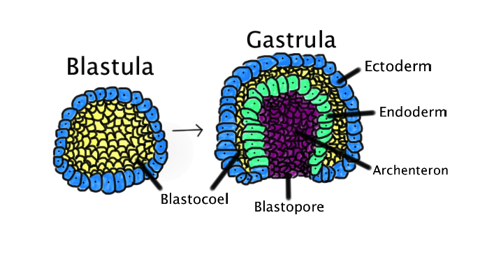
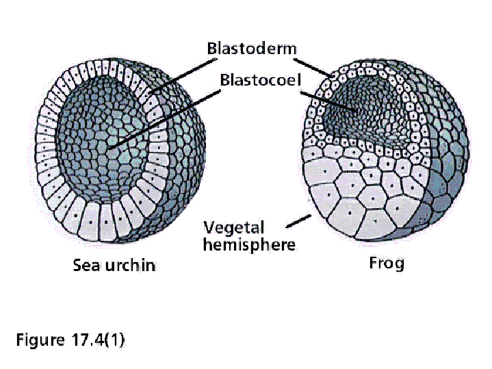
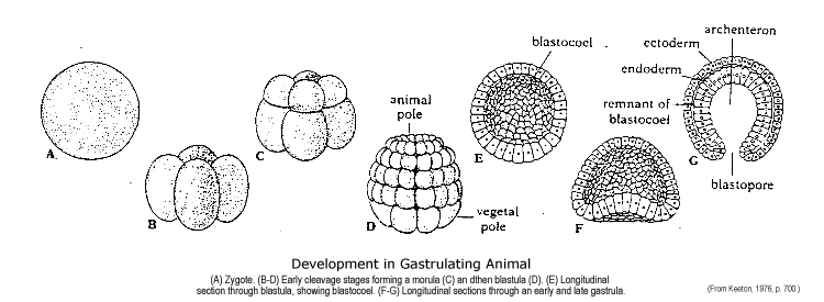
The blastula is a hollow sphere of cells, referred to as blastomeres, surrounding an inner fluid-filled cavity called the blastocoele formed during an early stage of. Blastula, hollow sphere of cells, or blastomeres, produced during the development of an embryo by repeated cleavage of a fertilized egg. The cells of the blastula form an epithelial (covering) layer, called the blastoderm, enclosing a fluid-filled cavity, the blastocoel.
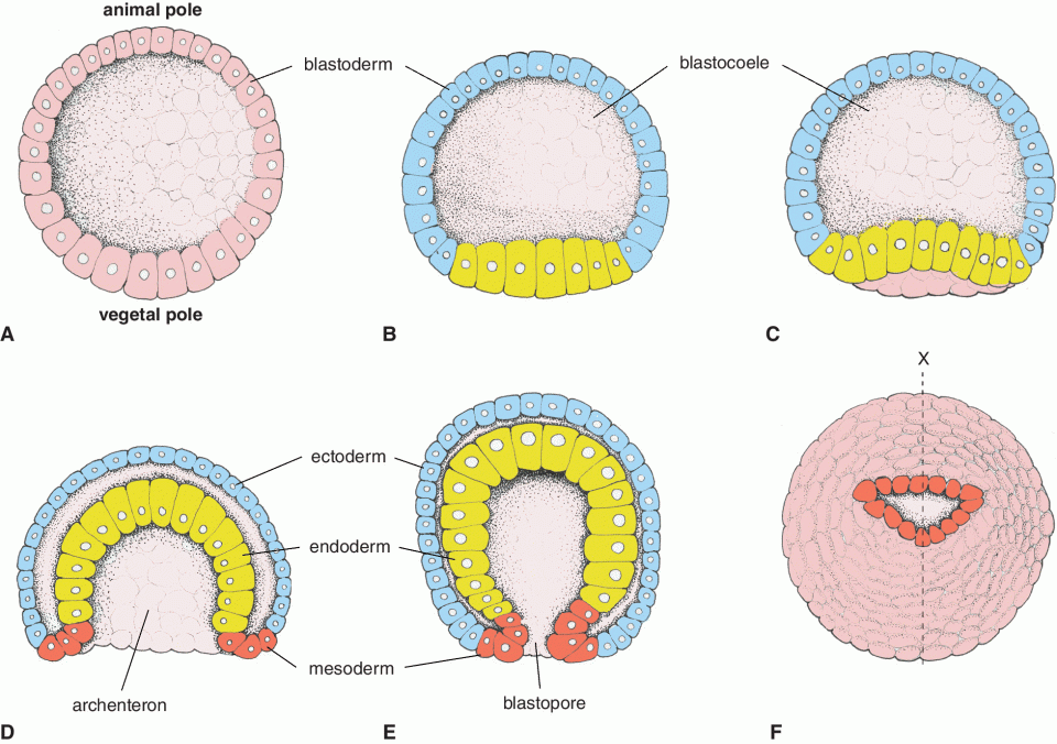
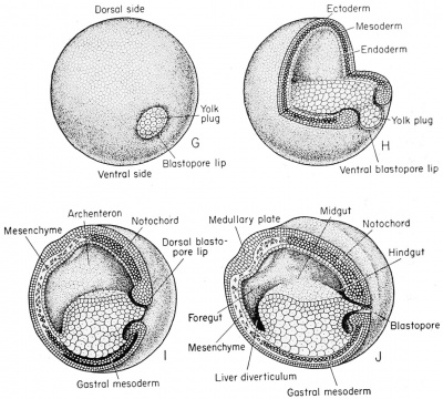
Blastula – embryo composed of many Blastomeres and a hollow center. Sea Urchin embryology: diagram Blastocoel – cavity of the blastula.The trophoblast cells on the outside of the blastula eventually became the placenta, while the inner cell mass inside of the blastula began to divide and form the developing embryo, you. Gastrulation is a phase early in the embryonic development of most animals, during which the single-layered blastula is reorganized into a multilayered structure known as the gastrula.
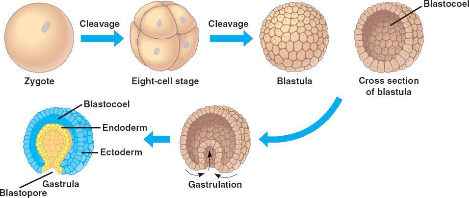
Prepared slide of whitefish blastula; Procedure. In Figure 6, identify the phase of mitosis and write the name of the phase below each diagram. The cells go in the appropriate temporal sequence through cell cycle and you are likely to use the same term multiple times.
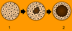
Blastula, hollow sphere of cells, or blastomeres, produced during the development of an embryo by repeated cleavage of a fertilized egg. The cells of the blastula form an epithelial (covering) layer, called the blastoderm, enclosing a fluid-filled cavity, the blastocoel. This fate map diagram of a Xenopus blastula shows cells whose fate is to become ectoderm in blue and green, cells whose fate is to become mesoderm in red, and cells whose fate is to become endoderm in yellow.
Notice that the cells that will become endoderm are NOT internal!Frog EmbryologyMetaphase Whitefish Blastula Cell
