
Download scientific diagram | Euplotes dragescoi nov. spec. after protargol impregnation.
(A, B) Ventral and dorsal views of the same specimen, arrow in A. A population of Euplotes collected from the mouths of the Rhône was assigned to the ..
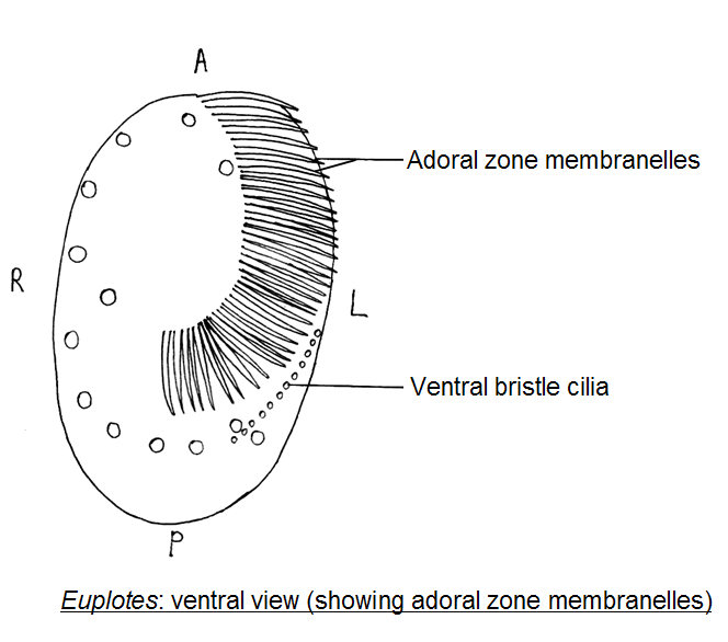
For interpretation of these figures also see the diagram in Figure Richard Allen’s collection of micrographs. Chapter Euplotes.
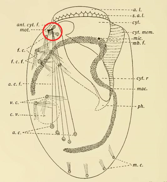
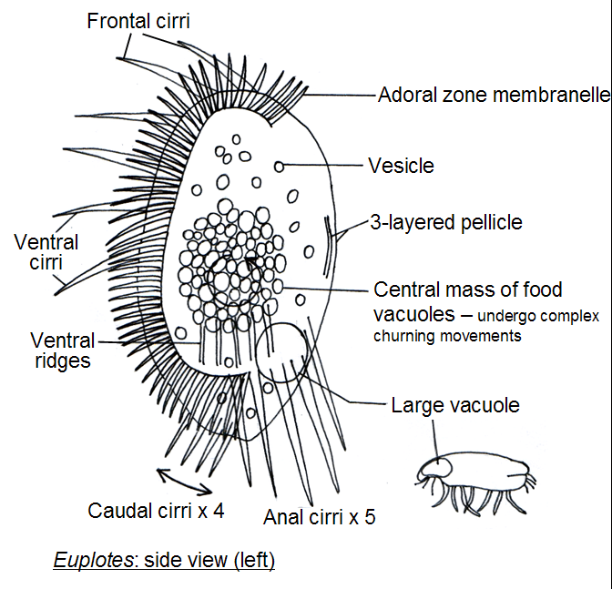
eup 13 — Fig 1: Euplotes sp. – sectioned tip to tip through cell’s widest part.
Download scientific diagram | Schematic drawings of Euplotes. (a) Ventral surface. There are five morphogenetic streaks along which the frontoventral (FVC ).
Download scientific diagram | Schematic drawings of Euplotes. (a) Ventral surface. There are five morphogenetic streaks along which the frontoventral (FVC ).Membrane control of ciliary activity in Euplotes c CRT *Vm Text-fig, i.
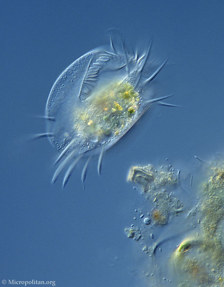
Diagram of apparatus. The specimen was held against the lower surface of the. Figure 1.
Identification of a TERT Gene Family in Euplotes crassus (A) Diagram of EcTERT-1, -2, and -3 coding regions. Asterisks denote the position of internal stop codons. Conserved RT and telomerase motifs, indicated in gray, were identified assuming that one or two +1 translational frameshifts occurred to generate full-length proteins.
Euplotes iliffei schematron.org: A new species of Euplotes (Ciliophora, Hypotrichida) from the marine caves of Bermuda. Bruce F.
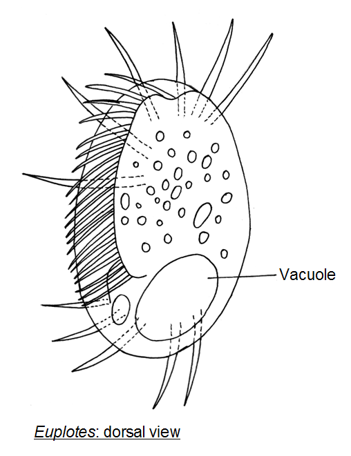
Hill Department of Biology, Georgetown University, Ink line diagram of the ventral aspect based on a protargol stained specimen. EUPLOTES ILIFFEI Ma.
Identification of a TERT Gene Family in Euplotes crassus (A) Diagram of EcTERT-1, -2, and -3 coding regions. Asterisks denote the position of internal stop codons. Conserved RT and telomerase motifs, indicated in gray, were identified assuming that one or two +1 translational frameshifts occurred to generate full-length proteins.
The Euplotes belong to the Phyllum Ciliophora. They are from µm long.
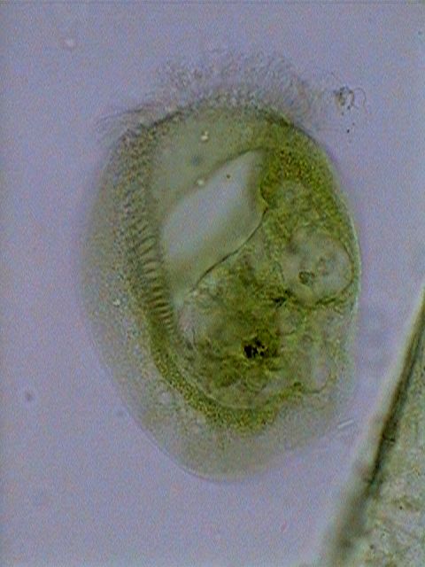
Euplotes is an interesting ciliate with a transparent body. It has large cilia that is tufted together to form cirri and a band-like macronucleus (the big backward “C” shown inside the body).Hypotrich – WikipediaHypotrich – Wikipedia
Paramecium Dividing