Water flea experiment from Microscopes for Schools. Observing daphnia pulex ( water flea) under a compound microscope.
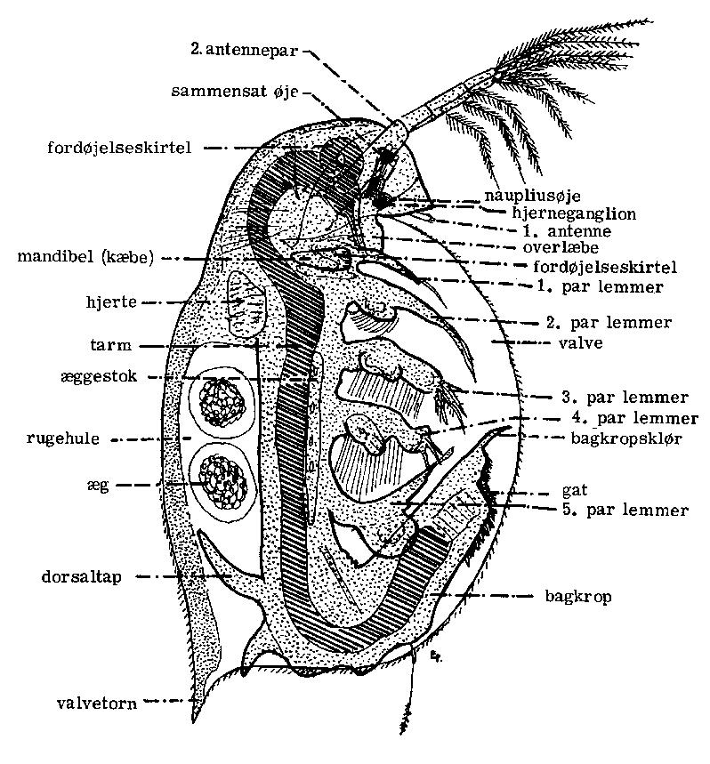
Daphnia swim by rapid downward strokes of the second large antenna, as illustrated in the diagram. The thoracic appendages are the leaf-like limbs that. Daphnia diagram of parts.

Below is an image of Daphnia that was captured using brightfield microscopy with a compound microscope at x magnification. Daphnia, a genus of small planktonic crustaceans, are –5 millimetres (– in) in length.
Daphnia are members of the order Cladocera, and are one of. Students observe daphnia with and without a magnifier. ..
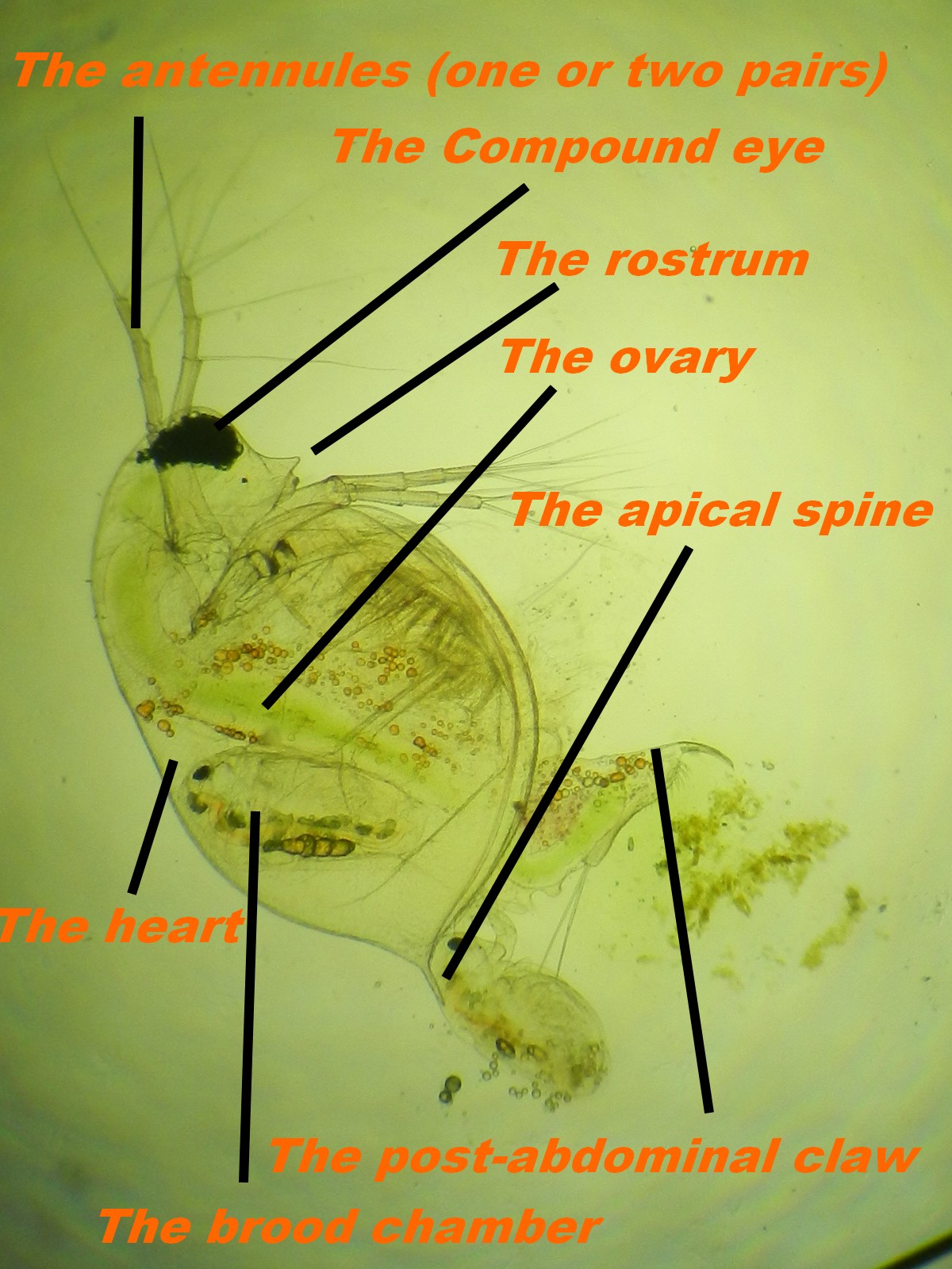
Point out the daphnia’s heart on the diagram, along the back the daphnia and label its body parts.Daphnia Body Parts STUDENT RESOURCE INFORMATION SHEET Esophagus Heart Food tube Eggs Babies are born live Carapace Anus (shell) Legs Filter hairs Antenna Mouth Eye Cells. Title: SEC04pg_indd Author: New Created Date.
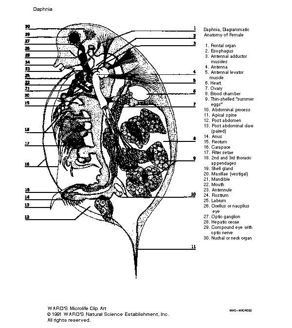
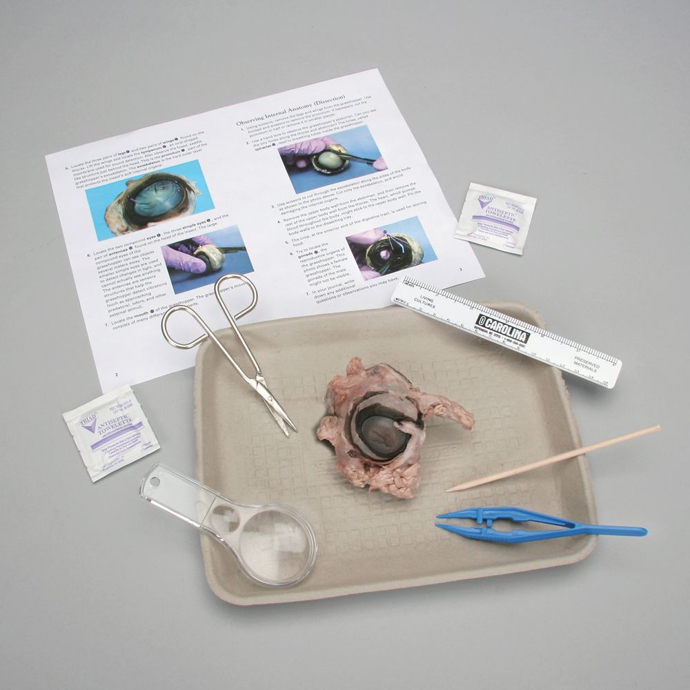
Labeled Organs Of Daphnia Magna Daphnia Magna Culture Kit, Living | Carolina. Labeled Organs Of Daphnia Magna Daphnia Magna Culture Kit, Living | Carolina – Labeled Organs Of Daphnia Magna.
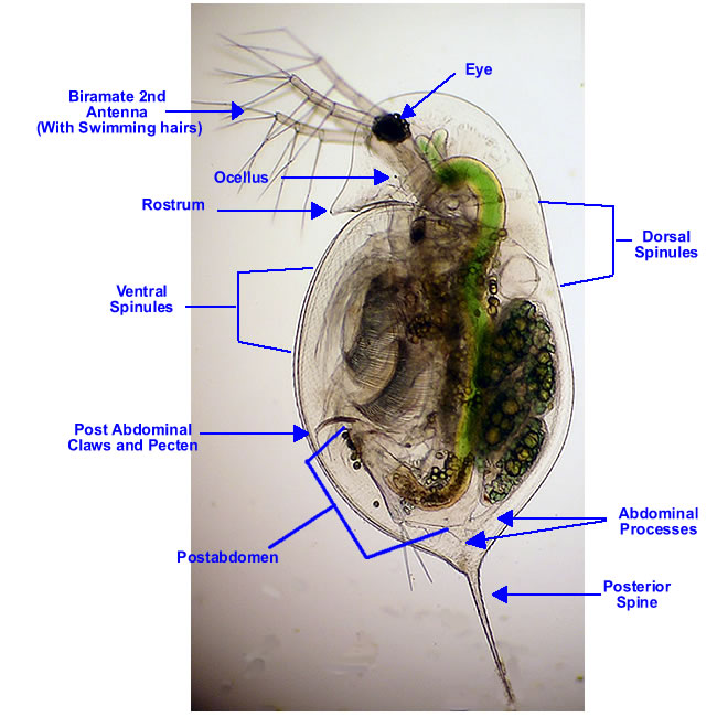
Back To Labeled Organs Of Daphnia Magna. 12 photos of the “Labeled Organs Of Daphnia Magna”. Labeled Organs Of Daphnia Magna Daphnia Magna Culture Kit, Living | Carolina photo, Labeled Organs Of Daphnia Magna Daphnia Magna Culture Kit, Living | Carolina image, Labeled Organs Of Daphnia Magna Daphnia Magna Culture Kit, Living | Carolina gallery.
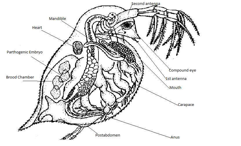
Human Skeleton With Veins Diagram. Human Digestive System Organs. Click on the options to the left to change the visible labels on this Daphnia.
To be able to use the Daphnia as a bioassay tool, they have to be familiar with normal Daphnia behavior and be somewhat familiar with Daphnia anatomy, see the Daphnia anatomy sheet labelled. 4. In groups have students look at the Daphnia within the beakers.Daphnia – WikipediaDaphnia Anatomy – A STUDY OF THE HEART
