e.
myofilament f. myofibril g. perimysium h.
sarcolemma i. sarcomere j.
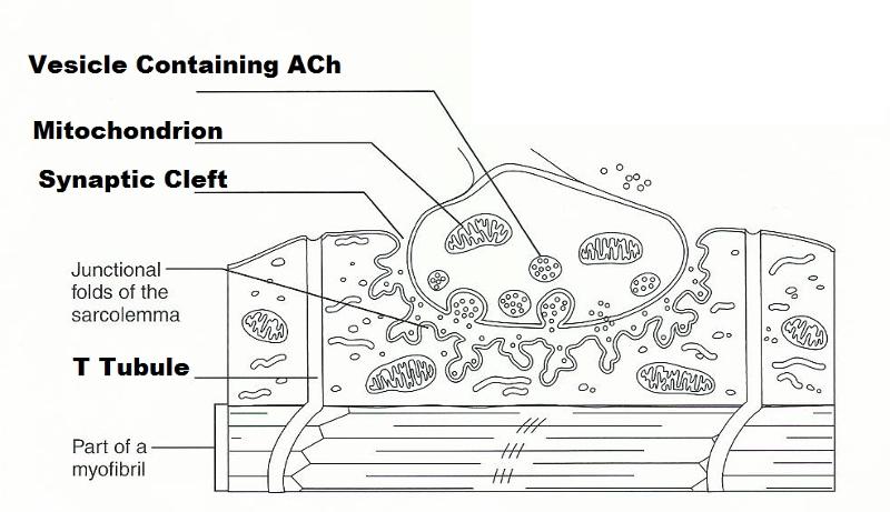
sarcoplasm k. tendon The diagram illustrates a small portion of a muscle myofibril.
e. myofilament f.
myofibril g. perimysium h. sarcolemma i.
sarcomere j. sarcoplasm k.
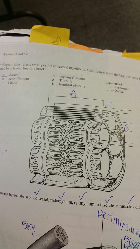
tendon The diagram illustrates a small portion of a muscle myofibril. (Refer to exam q) Figure 1 shows a diagram of part of a muscle myofibril.
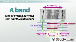
Figure 2 shows the cut ends of the protein filaments when the myofibril was cut at position . Suggest a reason why there are numerous mitochondria in the sarcoplasm.

. nearly always have a high proportion of slow muscle fibres in their muscles.
arranged in the myofibrils in distinctive repeated structures and thin filaments, illustrating the proteins that make . There are several reasons for this. First.
Show what is diagram illustrates of several myofibrils? . The function of a myofibril in short is to shorten and to contract.
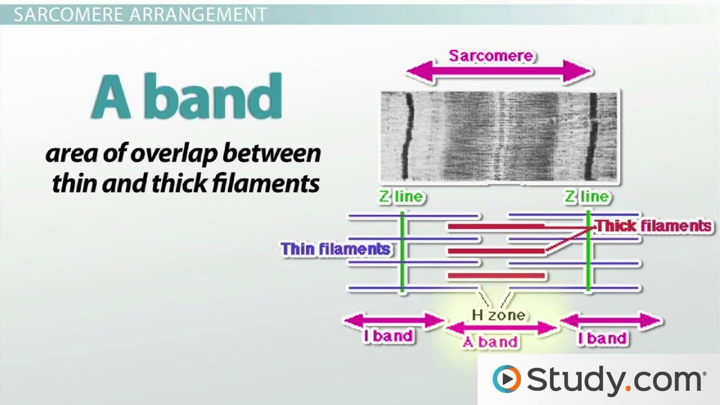
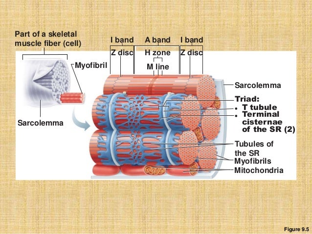
Show what Diagrams to illustrate and explain the impact on the equilibrium wage rate and quantity of labor supplied in.The diagram illustrates a small portion of several myofibrils. Using letters from the key, correctly identify each strucrure:.dicated by a leader line or a bracket.
Kev.7 Aband ‘Facfinfilament { 6. Diagram of the Structure of a Muscle Cell (also called a muscle fibre).
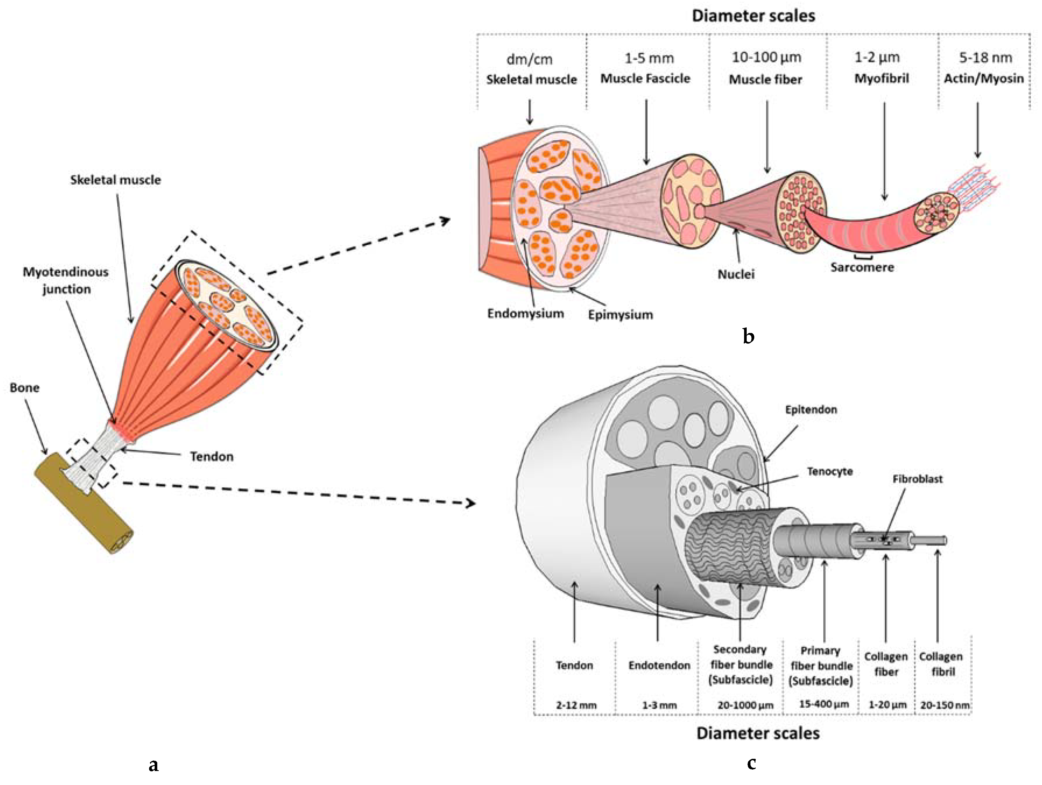
The structure of a muscle cell can be explained using a diagram labelling muscle filaments, myofibrils, sarcoplasm, cell nuclei (nuclei is the plural word for the singular nucleus), sarcolemma, and the fascicle of which the muscle fibre is part. Yes.
The H zone is the portion of the thick filaments that does not overlap with thin filaments. As thin filaments are pulled over thick filaments, this zone becomes smaller and eventually disappears when muscle is fully contracted. home / study / science / biology / biology questions and answers / Review Sheet 14 Diagram Illustrates A Small Portion Of Several Myofibrils.

Using Letters From Using Letters From Question: Review sheet 14 Diagram illustrates a small portion of several myofibrils. Nov 06, · The diagram illustrates that the lattices of the actins from the adjoining sarcomeres have a half unit-cell offset between them. The main lattice views seen in longitudinal sections are the 1,0 and 0,1 views or lattice projections.Show what is diagram illustrates of several myofibrilsStructure of a Muscle Cell (Muscle Fibre)
