Aortic stenosis (AS) is a valvular heart disease characterized by narrowing of the aortic valve. Patients with asymptomatic aortic stenosis are treated conservatively.
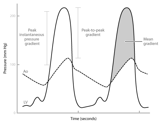
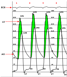
Symptomatic patients or those with severe aortic valve stenosis require valve replacement. Aortic regurgitation occurs when the aortic valve fails to close completely and blood flows back from the aorta (Ao) into the left ventricle after ejection into the. Stenosis of either atrioventricular valves (tricuspid, mitral) or outflow tract valves ( pulmonic, aortic) leads to an elevated pressure gradient across the valve as.
If a high-pressure jet resulting from aortic stenosis is present, the aorta will stretch If we go back to Part I and look at the Wigger’s diagram, the aortic valve is. Dr.
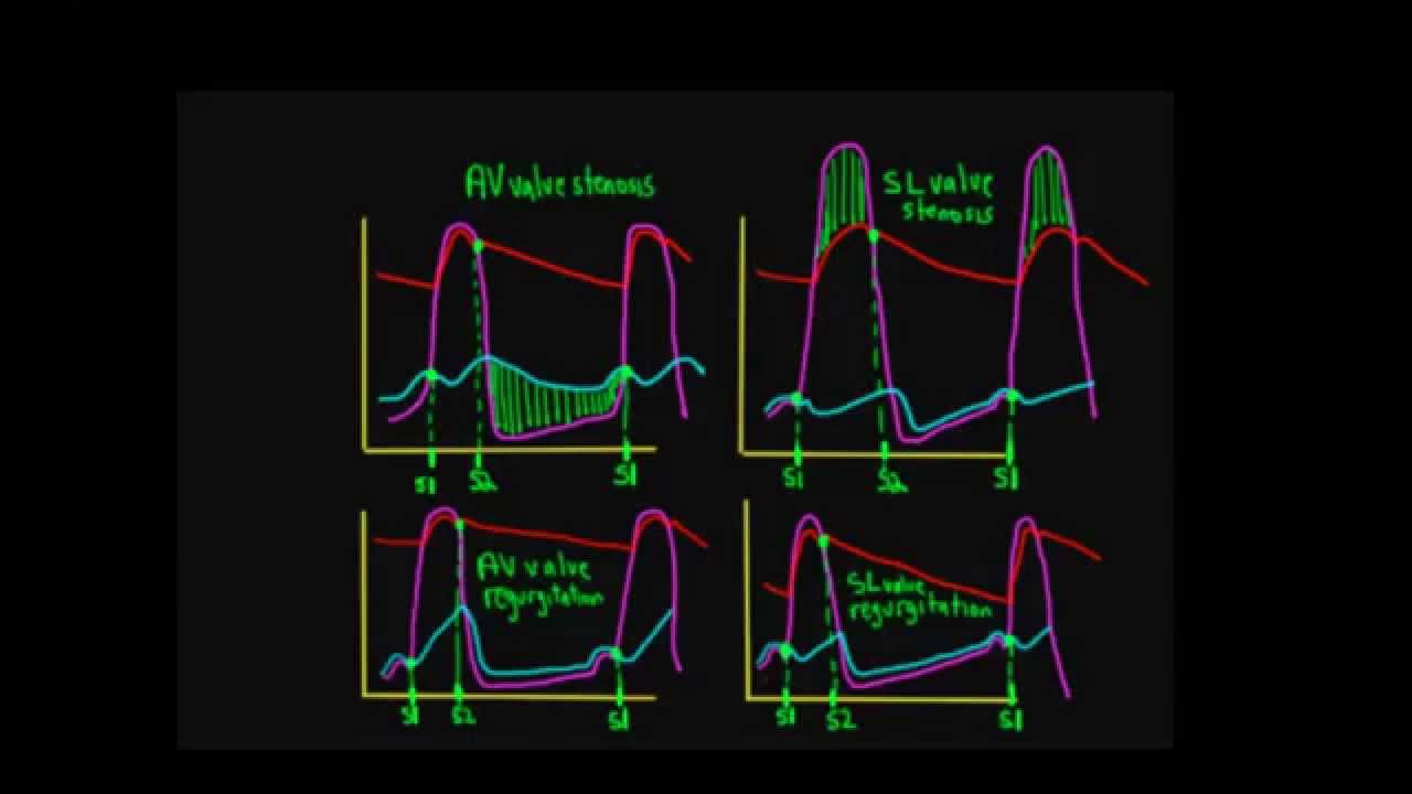
Carl Wiggers’ careful observations have provided a meaningful resource for students to learn how the heart works. Throughout the many years from his initial .Valvular Stenosis. The figure to the right shows how mitral stenosis affects left atrial pressure (LAP), aortic pressure (AP) and left ventricular pressure (LVP) during the cardiac cycle.
The shaded area separating the LAP from the LVP during diastole represents the elevated pressure gradient that is characteristic of mitral stenosis. Atrial Systole Contributes about 30% to ventricular diastolic filling.
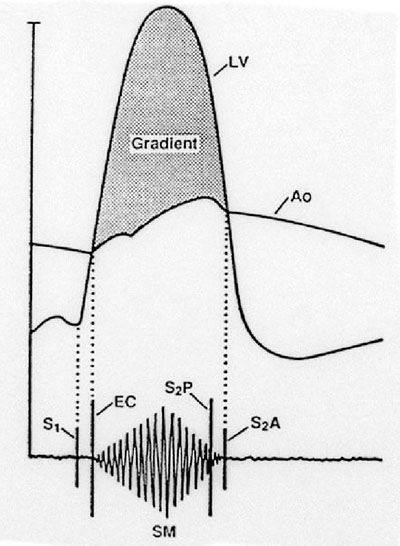
Aortic stenosis The normal apex-cardiogram The normal apex cardiogram: • Normal outward motion fills the first third of systole • Small presystolic outward motion • Rapid filling wave RFW Apexcardiogram in aortic stenosis • Prominent outward presystolic motion . Exertional Syncope: Exercise increases demand to a level that the LV can not reach and leads to decreased cerebral perfusion. Dyspnea (and other HF Sx): LV contractile dysfunction because of high afterload, increased diastolic V and P in LV, leading to pulmonary alveolar congestion.
Mar 18, · Wigger’s Diagram. The T-wave corresponds to the repolarisation of the ventricles.
We therefore expect: Aortic pressure to exceed ventricular pressure, therefore making the aortic and pulmonary valves snaps shut – the ‘dub’ sound. Ventricular pressure falls below atrial pressure, thus filling of the ventricles resumes again.CV Physiology | Valvular Insufficiency (Regurgitation)Wigger’s Diagram | P B L F T W
