When studying the anatomy of amphibians, it is essential to have a clear and precise representation of their internal systems. The internal organs of these creatures can be explored through a step-by-step breakdown, providing valuable insights into their biology. This detailed view helps students and researchers alike understand how the body parts function and interact within the organism.
Focus on the digestive system, which includes the mouth, esophagus, stomach, and intestines. The circulatory system is also important, involving the heart, arteries, and veins. Pay close attention to how these organs are connected and arranged within the body cavity, as this reflects the overall efficiency of the organism’s internal processes.
For a comprehensive understanding, it is recommended to examine the respiratory system, noting the location of the lungs and gills, which play distinct roles at different stages of the creature’s life cycle. Understanding the nervous system, including the brain and spinal cord, is equally important to grasp how the animal responds to stimuli and coordinates movement.
By thoroughly studying these systems, one gains a deeper appreciation for the evolutionary adaptations that enable survival in various environments. A well-executed internal view offers a structured approach to learning the complex workings of amphibians.
Detailed Overview of Internal Anatomy

When studying the internal structures of amphibians, focus on the essential organs like the heart, lungs, liver, and kidneys. The heart, typically three-chambered, is crucial for understanding circulatory processes. Observe its position and relationship with the blood vessels. Pay special attention to the two atria and one ventricle, noting the direction of blood flow between them.
The respiratory system involves both gills and lungs, with the latter developing after metamorphosis. The lungs are responsible for gas exchange, while the gills assist during earlier life stages. Trace the airway path from the mouth to the lungs and the connection to the trachea.
The digestive system, consisting of the mouth, esophagus, stomach, and intestines, plays a key role in nutrient processing. Examine how food moves through these organs and how nutrients are absorbed. The liver and pancreas support digestion by producing enzymes and bile necessary for breaking down food particles.
Examine the kidney’s role in filtration and waste excretion. It’s essential to locate the kidneys within the body cavity, as they help regulate fluid balance and maintain homeostasis. The urinary system also includes the urinary bladder, where waste is stored before excretion.
In the reproductive system, focus on the gonads and their connection to the urogenital ducts. In males, testes are found near the kidneys, while in females, ovaries are situated in a similar location. These organs produce gametes for reproduction and contribute to the species’ life cycle.
Be thorough in noting the nervous system’s structure, especially the spinal cord and brain. The central nervous system controls movement, sensory input, and reflex actions. The spinal cord is the main communication route between the brain and body, while the brain processes sensory data and coordinates activities.
Identification of Internal Organs in a Frog Dissection

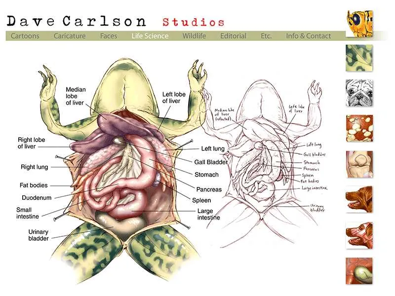
To identify the internal organs, first locate the abdominal cavity. Use scissors to make a midline incision, cutting through the body wall. The first structure visible is the liver, a large, dark-brown organ. It consists of three lobes: the left, right, and median lobes. Gently move the liver to expose the heart beneath it, a small, triangular structure located at the center of the chest cavity. The heart has three chambers: two atria and one ventricle.
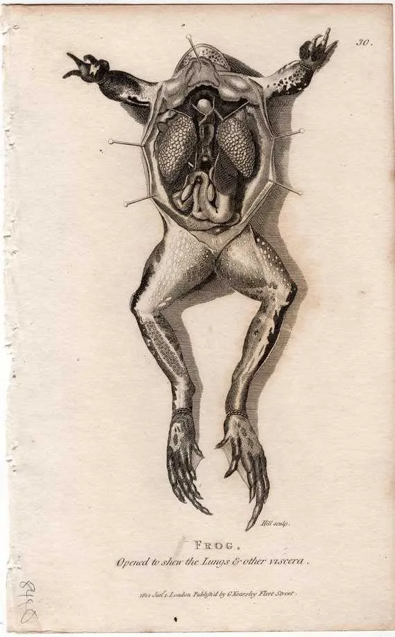
Next, find the lungs on either side of the heart. These are relatively small, spongy organs responsible for gas exchange. Below the heart, the intestines, a long, coiled tube, are visible. Trace the small intestine to its junction with the large intestine, which is broader and shorter. At the posterior end of the large intestine, locate the cloaca, a common chamber for excretion and reproduction.
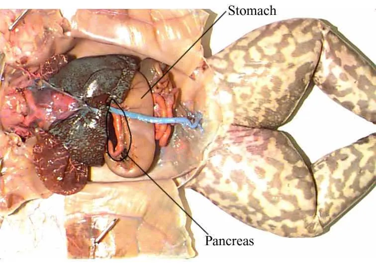
The stomach lies on the left side of the body, just beneath the liver, and is connected to the small intestine. It has a J-shaped curve. Identify the pancreas, a small, glandular structure near the junction of the stomach and small intestine, which produces digestive enzymes.
Near the intestines, find the kidneys, located along the dorsal side of the body. These are bean-shaped organs responsible for excretion and osmoregulation. The urinary bladder lies between the kidneys and is used for storing waste before excretion.
Finally, observe the reproductive organs. In males, the testes are located near the kidneys, while females have large, oval-shaped ovaries. These are usually filled with eggs and can be identified by their texture.
Step-by-Step Guide for Drawing a Frog Dissection Diagram

Start by choosing a high-quality reference image of the specimen. Make sure the image clearly shows the internal organs and structures you wish to illustrate.
- Sketch the Outline: Begin with a light pencil sketch of the specimen’s body. Focus on capturing the overall shape and proportions. Pay close attention to the placement of limbs and the midline of the torso.
- Label Key Structures: Mark the general locations of the main organs, such as the heart, liver, lungs, and digestive system. This helps guide the detailed drawing in later steps.
- Draw the Internal Organs: Work systematically, drawing each organ with accuracy. For instance, outline the heart, intestines, and other systems while maintaining proper scale and relative positions.
- Highlight Anatomical Details: Use finer lines to represent the smaller structures, such as blood vessels, nerves, and ducts. Be sure to emphasize the textures of the organs where necessary.
- Use Shading: Lightly shade the organs to give them depth and dimension. Apply darker shading on areas of the body that are farther from the light source.
- Label Every Structure: Place clear labels next to each organ and feature. Use a consistent font style and ensure that each label is easy to read.
- Refine the Drawing: Once the basic structures are in place, go over your lines with a darker pencil or pen. Make sure all details are clean and sharp.
- Finalize the Diagram: Review the drawing for accuracy. Double-check the labeling, proportions, and any anatomical features that need adjustments.
Ensure that the diagram is neat, with all details visible and properly labeled for educational use.
Common Mistakes When Labeling a Frog Dissection Diagram
One common error is incorrectly labeling the organs. For example, confusing the liver and pancreas is frequent, as both are located in the abdominal cavity and share a similar appearance. The liver is larger, more triangular, and located near the top, while the pancreas is smaller and positioned under the stomach.
Another mistake is failing to identify structures with similar names, such as the small and large intestines. The small intestine is coiled and thin, while the large intestine is broader and shaped like a U, ending at the cloaca. Pay attention to their size and shape differences when labeling.
Omitting or misplacing the heart is a frequent issue. The heart, consisting of two atria and one ventricle, is located at the top of the body cavity. It’s important not to confuse it with the lungs, which are located on either side of the heart.
Incorrect placement of the kidneys is another mistake. These organs lie along the dorsal side of the body cavity, but they should not be confused with the reproductive organs, which are also positioned near the same region.
Finally, failing to properly position labels next to the structures can cause confusion. Labels should be clearly placed near the corresponding organ, ensuring they do not overlap or obscure other vital parts, such as the digestive tract or circulatory system.