When studying the structure of a steed, it’s crucial to familiarize yourself with the key segments that make up its physical form. Understanding the placement and function of each region aids in better care, training, and overall management. Start by focusing on the head, where the sensory organs are located, including the eyes, ears, and nostrils, each serving vital roles in communication and awareness of surroundings.
Next, pay close attention to the neck and spine, as they support both movement and flexibility. The cervical vertebrae allow for precise head positioning, while the back plays a role in weight distribution, particularly when carrying a rider or pulling loads. The limb structure, especially the legs, is also fundamental. The joints, muscles, and hooves work in unison to ensure efficient movement, with the hooves requiring particular attention for health and maintenance.
For optimal performance and longevity, it is essential to understand how each system interacts. From the digestive tract, responsible for processing nutrients, to the circulatory system, which ensures oxygen and blood flow throughout, every part contributes to the overall well-being. Knowledge of these systems ensures that caretakers can maintain the steed’s health and maximize its potential.
Understanding the Anatomical Structure of a Steed
To effectively care for or study an equine, it’s crucial to familiarize yourself with its key body regions. Below are the main areas to focus on:
- Head: Includes the eyes, ears, nostrils, mouth, and the jaw. Ensuring proper dental care and eye health is essential for overall well-being.
- Neck: Important for posture and movement. Pay attention to stiffness or sensitivity, as these can affect the animal’s flexibility and range of motion.
- Back: Vital for carrying weight and absorbing shock during movement. Regular inspections for signs of discomfort or misalignment are advised.
- Chest: Houses the heart and lungs. A healthy chest area ensures optimal lung capacity for breathing and overall stamina.
- Flanks: Located just in front of the hind legs, these areas can indicate the overall health of the animal through muscle tone and skin condition.
- Hindquarters: This section supports the animal’s movement, including powerful strides and agility. Watch for lameness or uneven gait.
- Legs: Consist of several segments including the fetlock, cannon, and pastern. Regular checking for cuts, bruises, or swelling is crucial to avoid lameness.
- Tail: Primarily used for communication and balance. Its condition reflects the animal’s general health, especially in the case of skin issues or parasites.
By regularly checking these zones, you can ensure that any early signs of discomfort or disease are promptly addressed, promoting longevity and optimal health.
Understanding the Skeletal Structure of a Horse
The equine skeletal system consists of approximately 205 bones, which are categorized into several regions: the head, neck, trunk, limbs, and tail. Key elements include the cervical vertebrae, thoracic ribs, and limb bones. The back is supported by 18 vertebrae, with 6 in the neck, 13 in the chest, and 5 in the lumbar region. The pelvis is made up of three fused bones–ilium, ischium, and pubis–providing the foundation for rear limb attachment.
The forelimbs lack a bony connection to the trunk, relying on the scapula and muscles for stability. The humerus, radius, and ulna connect the shoulder to the elbow and wrist, with the carpal bones acting as shock absorbers during movement. The metacarpals form the legs, and the coffin bone supports the hoof structure.
Muscular development is critical for supporting the skeletal framework. Proper muscle strength prevents bone stress and contributes to fluid, balanced motion, especially during high-impact activities like running and jumping. The alignment of the joints plays a major role in the overall posture and functionality of the entire organism.
Key Muscles in Equine Anatomy and Their Functions

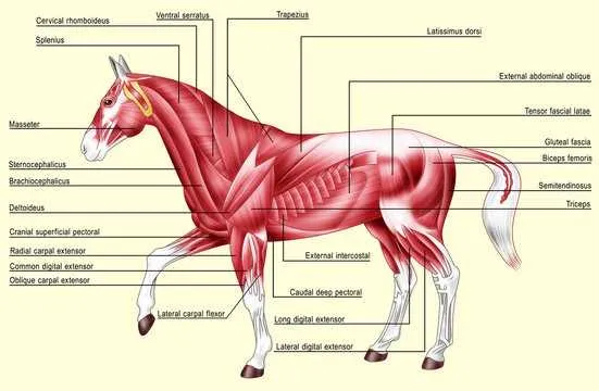
Trapezius: Located in the upper shoulder and neck, the trapezius plays a critical role in elevating and stabilizing the front limbs. It also assists with lateral neck movement, which is essential during various gaits and maneuvers.
Latissimus Dorsi: This large muscle is responsible for the movement of the forelimb, particularly in activities such as galloping. It also stabilizes the back and aids in overall posture during physical exertion.
Gluteus Medius: Positioned at the top of the hindquarters, this muscle is pivotal in propelling the animal forward during running or jumping. It provides thrust and helps maintain balance, especially when the body is in motion.
Quadriceps Femoris: Essential for locomotion, the quadriceps are involved in extending the hind limb and providing power during the push-off phase in a canter or sprint. These muscles are also key for stopping and decelerating movements.
Gastrocnemius: Known as the calf muscle, it assists in flexing the lower leg and is particularly active when the animal is running or jumping. It also contributes to the suspension phase during gaits like the trot and gallop.
Semitendinosus: Located in the hind limb, this muscle works to extend the hip and flex the stifle. It’s crucial for propulsion during the stride and helps in stabilizing the rear end during movement.
Deltoid: Positioned on the shoulder, the deltoid helps in lifting and rotating the front limbs, essential for effective turning and control during complex maneuvers.
Rhomboideus: This muscle aids in retracting the scapula, playing a significant role in maintaining the positioning of the forelimbs during various forms of movement.
Brachiocephalicus: This muscle spans from the shoulder to the neck, helping with forward movement of the forelimb and flexion of the neck. It also assists in head and neck posture during motion.
How Hooves Impact Performance and Health
Regular trimming and proper hoof care are essential for optimal athletic performance. Hooves should be checked every 6-8 weeks to prevent overgrowth, which can lead to uneven pressure distribution and discomfort. Inadequate hoof maintenance can cause lameness, poor gait, and difficulty in maintaining speed or stamina.
The balance of the hoof plays a crucial role in shock absorption. An imbalance may result in joint strain and long-term wear on muscles. Corrective shoeing is necessary if the natural hoof structure does not provide adequate support, especially in performance animals involved in rigorous activities such as jumping or racing.
Overly dry or moist hooves can lead to cracks or infections. Maintaining a consistent moisture level and applying hoof conditioners as needed prevents brittleness, especially in colder months. Additionally, ensuring proper footing in training and competition environments minimizes the risk of injury.
Hoof health is directly linked to overall well-being. A malnourished diet, especially one deficient in biotin and essential fatty acids, can negatively affect hoof integrity. It is important to include a balanced nutritional plan that supports healthy hoof growth to avoid frequent lameness or slow recovery from injuries.
Frequent inspection and timely intervention are key to preventing more severe complications. Addressing minor issues early can save time and cost, ensuring the animal’s longevity and maintaining peak performance levels over time.