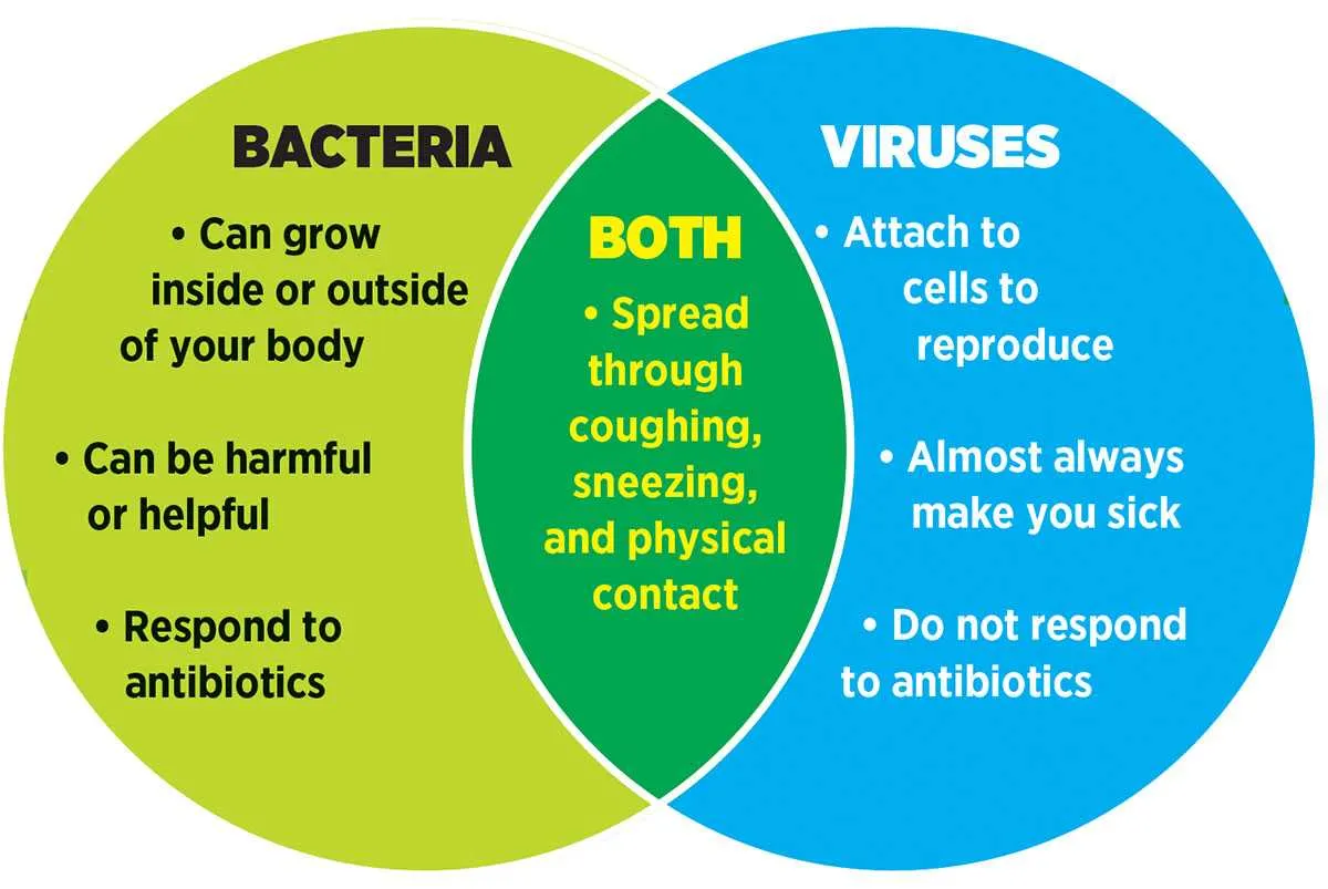
Use side-by-side charts to pinpoint distinctions and overlaps between unicellular organisms and infectious particles. These visuals help clarify size differences–one typically ranges from 0.2 to 2 micrometers, while the other measures in nanometers, often around 20–300 nm. The former possesses cell walls, ribosomes, and autonomous metabolic systems. The latter depends entirely on host cells for replication.
Identify shared traits to understand why both can trigger illness yet require different treatments. For instance, both can mutate rapidly and adapt to environmental pressures, but only one responds to antibiotics. The other demands targeted antiviral approaches or vaccines. Use colored intersections in schematics to emphasize commonalities like genetic material and capacity for rapid reproduction.
Highlight transmission modes to reinforce key preventive measures. One type spreads via contaminated surfaces, direct contact, or food, while the other often disperses through airborne droplets or vectors. Visual aids should clearly separate these mechanisms to support accurate public health messaging.
Comparison of Microbial and Viral Characteristics
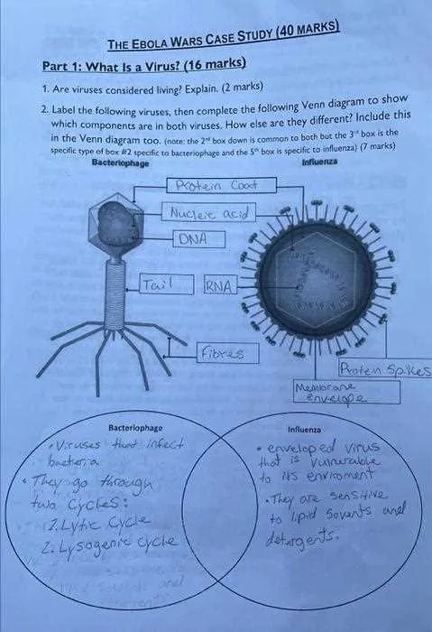
Start by grouping unicellular prokaryotes based on traits like cell walls, ribosomes, and independent reproduction. Contrast these with pathogens that require host cells, highlighting absence of organelles, genetic material encapsulated in protein coats, and host-specific replication.
List shared traits such as genetic code (DNA or RNA), ability to mutate, and transmission through air, surfaces, or fluids. Emphasize the overlap only in functional behavior–not in structure or autonomy.
For educational visuals, divide attributes into three sectors: unique to free-living microbes (e.g., antibiotic susceptibility, binary fission), exclusive to parasitic agents (e.g., capsid, reliance on host enzymes), and shared features (e.g., evolution, adaptation to environment).
Avoid merging traits like motility or metabolic processes, since only cellular organisms possess active mechanisms. Include examples such as *E. coli* for cellular forms and Influenza for host-dependent entities.
Use color-coded charts to enhance distinction: blue for self-replicating units, red for obligate intracellular invaders, and purple for mutual properties. Prioritize traits influencing treatment options, such as resistance mechanisms and infection pathways.
How to Visually Distinguish Structural Traits of Microbial Entities
Start by identifying the presence of a cell wall and internal organelles. Cellular microbes exhibit a rigid outer shell composed of peptidoglycan and possess ribosomes, cytoplasm, and sometimes plasmids. These components indicate a complete metabolic system.
In contrast, non-cellular agents lack such internal machinery. Look for a protein coat (capsid) surrounding nucleic acid, often either DNA or RNA, but never both. Some have an outer lipid envelope derived from host membranes–absent in simpler forms.
Size is a key clue: cellular forms typically measure between 0.2–2.0 micrometers, while acellular ones range from 20–300 nanometers. Electron microscopy reveals this disparity clearly.
Shape also helps: rod, spiral, or spherical profiles suggest self-sustaining units, while geometric or icosahedral structures point to parasitic particles.
Another marker is self-movement. Flagella or pili are exclusive to living microbial cells, aiding mobility or conjugation. Acellular forms rely entirely on hosts and exhibit no motility structures.
What shared characteristics should be placed in the overlapping section
- Microscopic size – both entities are invisible to the naked eye and require magnification for observation.
- Contain genetic material – each possesses either DNA or RNA as the blueprint for replication and function.
- Capable of evolution – undergo mutations and natural selection, adapting to environmental pressures over time.
- Cause infectious diseases – both can trigger illness in humans, animals, or plants through cellular disruption.
- Spread via transmission – dispersed through air, water, contact, or vectors like insects or animals.
- Target specific hosts – often exhibit host specificity due to surface molecule compatibility.
- Reproduce quickly – under favorable conditions, replication occurs at rapid rates, enhancing survival.
When to use a Venn diagram in microbiology education or research

Apply overlapping schema during comparative analysis of prokaryotic versus viral life forms, especially when teaching genomic organization, replication methods, or immune evasion tactics. Such visual juxtaposition supports immediate recognition of both exclusive and shared biological features.
Instruct undergraduate cohorts using intersecting sets when distinguishing cell structure, metabolic capability, or environmental adaptation. This enhances conceptual clarity and minimizes rote memorization.
Utilize dual-set models during literature reviews or meta-analyses to categorize gene expression profiles, host interactions, or resistance mechanisms across microbial agents and phage entities. This improves pattern detection and hypothesis formulation.
Implement during curriculum design to integrate topics like horizontal gene transfer or pathogenic pathways across different microbial domains. Such configurations aid in aligning learning objectives with cognitive skill development.