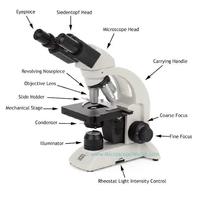
To fully understand how an optical instrument functions, it’s crucial to identify and comprehend the key elements that contribute to its operation. A thorough analysis reveals a range of critical structures, each serving a unique role in magnifying minute objects for clearer observation. Ensuring familiarity with these components enables efficient use and troubleshooting.
Objective lenses are fundamental in determining the degree of magnification. Positioned near the sample, these lenses vary in power, typically ranging from 4x to 100x. The higher the magnification, the closer the distance between the lens and the specimen.
Eyepieces allow the viewer to observe the enlarged image formed by the lenses. These optical elements often include a reticle for measuring the size of observed items. It’s essential to choose an eyepiece with appropriate magnification for the task at hand, ensuring both clarity and precision.
For focusing on specimens, the coarse and fine focus knobs provide necessary adjustments. The coarse knob moves the platform vertically, enabling a rough focus, while the fine knob refines the focus to achieve maximum clarity. Precision in these controls directly impacts image sharpness.
Illumination systems, such as mirrors or built-in lights, enhance visibility by illuminating the sample. Proper lighting is crucial to obtaining high-quality images, particularly when working with transparent specimens that require backlighting.
Finally, a solid stage serves as the stable platform on which the sample is placed. Depending on the type of instrument, stages may feature mechanical movements for precise positioning of the sample, ensuring that the area of interest is always in the center of the field of view.
Understanding Key Components of a Microscope
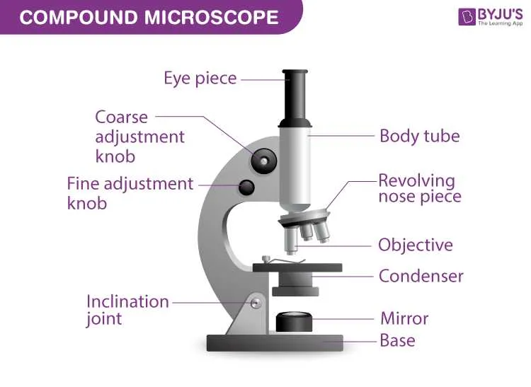
Focus on the ocular lens when viewing an object under magnification; this allows you to see the image clearly. The objective lenses, typically ranging from low to high magnification, determine the level of detail visible. Adjust the stage to position the specimen correctly. Use the coarse adjustment knob for rough focusing and the fine adjustment for precision.
The light source illuminates the specimen, making it visible. A diaphragm controls the amount of light, ensuring optimal contrast and clarity. The arm and base provide structural support, holding everything in place while allowing easy manipulation during observations.
For high-powered imaging, ensure proper alignment of the condenser lens, which focuses light onto the specimen. A rotating nosepiece enables quick switching between objectives without moving the sample.
Regular cleaning of lenses ensures clear images. Use lens paper and gentle motion to avoid scratches. Ensure proper storage to prevent damage to delicate components.
Identifying Key Components of a Microscope: Lens, Stage, and Focus Mechanism
Objective lens is crucial for high-resolution imaging. Choose a lens with appropriate magnification to match your observation needs. For clear views, ensure the objective lens is clean and free of debris.
Stage supports slides. Ensure it is securely locked in place to prevent any shift during use. For accurate positioning, use the mechanical stage controls to move the specimen precisely. If there is a stage clip, check that it’s functioning to hold slides firmly.
Focus system is vital for sharp, clear images. Begin with coarse focus adjustment to locate the specimen, then use fine focus for detailed examination. Keep the lens and specimen at an optimal distance for clarity–adjust until the image appears crisp.
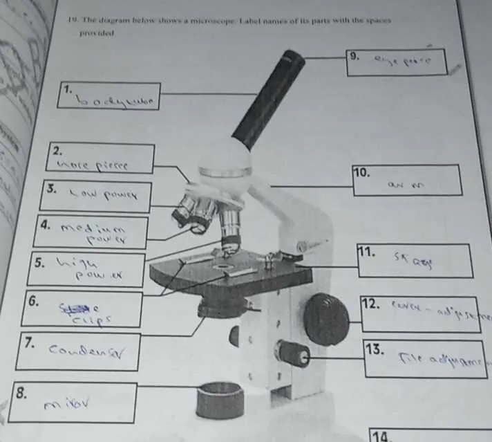
How to Read a Microscope Diagram: Understanding Labels and Functions

Start with the eyepiece: identify the lens at the top through which observation occurs. Note its magnification, typically 10x.
Locate the objective tubes: these hold multiple lenses with varying powers (4x, 10x, 40x). Understand which lens is in position for current viewing.
Check the arm: this vertical support connects the base to the top structure. It’s used for safe handling and stabilizing the optical setup.
Identify the stage: this flat platform supports the sample. It usually includes clips to secure slides in place.
Find the coarse and fine focus knobs: larger knob adjusts overall clarity; smaller one sharpens the image without major shifts.
Observe the light source: located beneath the specimen platform, it illuminates the slide from below. Some versions use mirrors instead of built-in bulbs.
Note the diaphragm: this rotating disc under the platform controls light intensity. Adjust for contrast based on sample transparency.
Look for the nosepiece: a rotating turret allowing quick lens changes. Align properly to ensure lens clicks into viewing position.
Tip: Always match label numbers to these features before interpretation. Misreading a label leads to incorrect understanding of optical function.
Common Misconceptions About Microscope Components and How to Avoid Them

Never rotate the coarse adjustment knob while using high magnification. Doing so can crack the slide or damage the objective. Always switch to fine focus once the longer lens is in place.
- Eyepiece magnification is often misunderstood. Many assume total magnification equals only the ocular’s power. In fact, it’s the product of ocular and objective strengths. For example, 10× ocular with 40× lens gives 400× total.
- Illumination control is frequently neglected. Users often ignore the diaphragm, relying solely on the built-in light. Adjust aperture size to enhance contrast, especially for transparent specimens.
- Cleaning lenses with rough cloths leads to scratches. Use lens paper or microfiber. Never apply cleaning fluid directly–moisten the paper instead.
- Stage clips are not meant to force slides into place. Improper use causes cracking. Align carefully and secure without pressure.
- The condenser is often underutilized. Raise it close to the stage when using high-power optics to improve resolution and brightness. Lower it slightly for low-power observations.
- Cover slips aren’t optional. Some skip them during wet mount prep, causing air bubbles or contact with objectives. Always use a properly placed coverslip.
Consistently misidentifying objectives as interchangeable across devices can lead to poor imaging. Each lens is calibrated for specific optical paths. Stick to original sets unless professionally matched replacements are available.