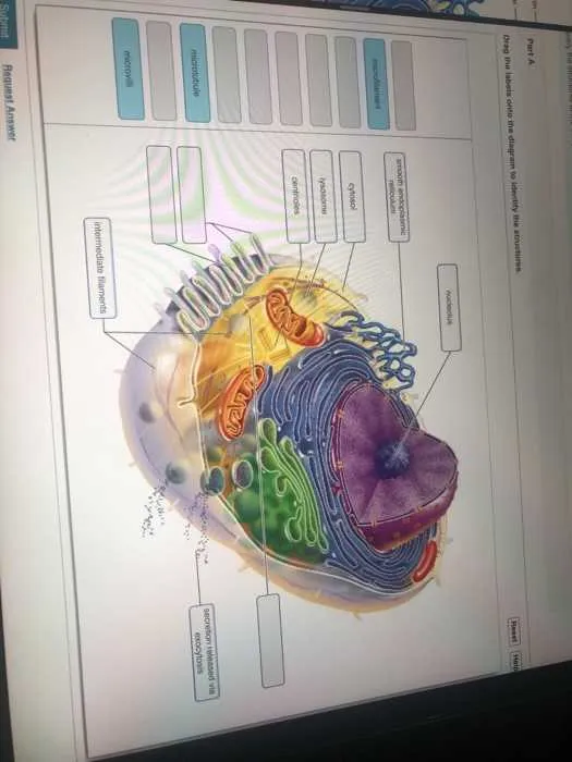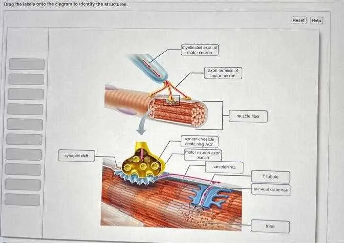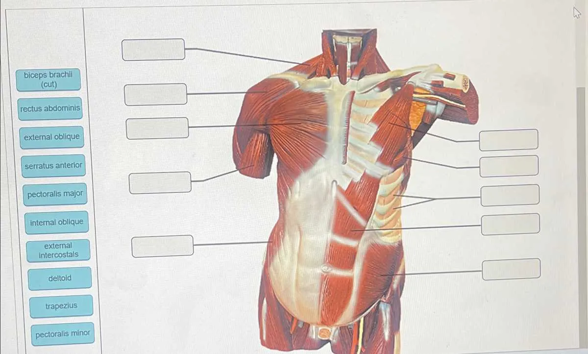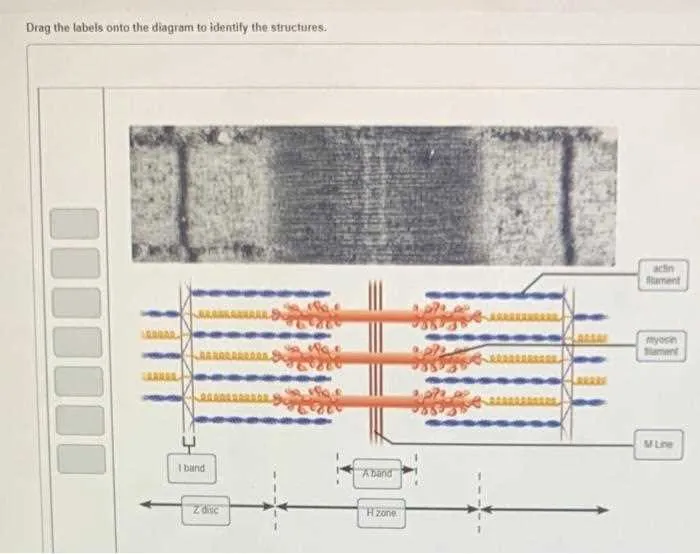
Begin by analyzing each element depicted in the image. Focus on shape, location, and surrounding context to match each term precisely. Incorrect placement often results from superficial observation; examine anatomical borders and spatial relationships carefully.
Prioritize distinctive features. For example, tubular structures suggest vessels or ducts, while layered patterns typically indicate tissue groupings. Matching curvature, thickness, and branching patterns can also help determine the correct terminology for each component.
Use existing references or comparative charts to validate uncertain matches. If the image presents a biological system, such as nervous or circulatory, consult cross-sectional diagrams to reinforce placement accuracy. Double-check any ambiguous zones before proceeding to the next item.
Maintain logical flow–start from one side and proceed systematically. This approach reduces confusion and ensures consistency. Avoid random assignments; every term should correspond to visual and functional logic based on observable characteristics.
Precise Matching for Anatomical Components

Begin by pinpointing each biological feature based on spatial orientation and relative size. For example, when analyzing a neural cross-section, locate the central canal by identifying the smallest circular region surrounded by gray matter. Adjacent to this, the anterior horns can be recognized by their broader, curved extensions pointing forward.
Associate terminologies with visual indicators. For instance, when working with cardiovascular illustrations, connect “left atrium” to the chamber receiving oxygen-rich blood from pulmonary veins, situated posteriorly and superiorly. Meanwhile, align “right ventricle” to the muscular structure projecting blood toward pulmonary circulation.
Utilize symmetry and alignment as clues. In histological slides, such as epithelial layers, match terms to the basal region by identifying denser cell arrangements near connective tissue interfaces. Contrast this with the apical surface, where cells face open lumens or external environments.
Confirm associations through contextual cues. In skeletal mappings, relate names to bone morphology–such as placing “scapula” over the flat, triangular plate connected to the humerus and clavicle–ensuring anatomical accuracy through proportion and position.
How to Match Labels with Anatomical Features

Begin by locating distinct landmarks such as bones, organs, or muscle groups; these serve as fixed points of reference. Align each term with the corresponding shape, texture, or position in the illustration. For instance, pair “femur” with the longest bone in the upper leg region and “liver” with the large, lobed mass in the upper right abdominal quadrant.
Use directional cues like superior, inferior, medial, and lateral to refine placements. If a term suggests function (e.g., “respiratory structure”), associate it with organs like the trachea or lungs based on their role in airflow.
Double-check surrounding context–adjacent tissues or systems often provide clues. For example, if one part connects to a known organ or vessel, it’s likely part of that system. Match connective tissues, nerves, or vessels by tracing their paths between landmarks.
Validate accuracy by comparing bilateral symmetry where applicable. Inconsistent pairing on mirrored structures typically indicates a mismatch.
Common Mistakes When Labeling Diagrams

Always align text precisely with the relevant anatomical or structural part to avoid misinterpretation.
- Incorrect positioning: Placing annotations too far from the feature leads to confusion. Use leader lines or proximity for clarity.
- Overlapping terms: Avoid stacking names or letting them cross each other. Maintain spacing to preserve legibility.
- Inconsistent terminology: Mixing scientific and common terms in one illustration disrupts understanding. Choose one system and stick to it.
- Ambiguous pointers: Arrows or lines should terminate exactly on the intended region. Vague or floating indicators reduce accuracy.
- Repetitive descriptors: Don’t reuse the same word for different components. Each item must be uniquely defined.
- Neglecting scale or orientation: If directional indicators or magnification scales are missing, spatial relationships become unclear.
- Typographical errors: Misspellings or inconsistent capitalization reduce credibility and may result in incorrect comprehension.
- Visual clutter: Too many elements crammed in a small space obscure the core message. Simplify whenever possible.
To ensure accuracy, double-check terminology against reliable references, and validate placement with subject-specific criteria.
Tips for Practicing Diagram Labeling in Study Tools

Focus on segmenting complex images into smaller sections. Concentrate on one area at a time to improve memorization and reduce overwhelm. This approach allows for gradual mastery of each part before moving on.
Utilize mnemonic devices to associate terms with visual elements. Creating memorable connections between names and locations enhances recall during practice and testing scenarios.
Regularly time your practice sessions to build speed without sacrificing accuracy. Short, frequent intervals encourage retention more effectively than longer, infrequent attempts.
Incorporate varied study materials such as flashcards and quizzes alongside interactive exercises. This multimodal exposure reinforces comprehension and aids in recognizing patterns across different representations.
Review incorrect placements immediately. Analyzing mistakes and understanding why errors occur helps prevent repetition and deepens understanding of spatial relationships.