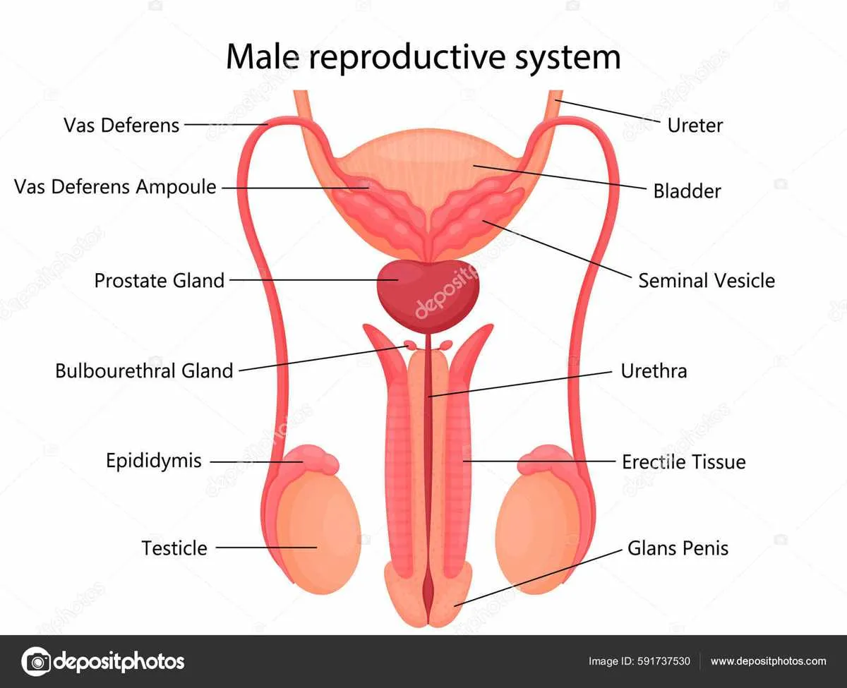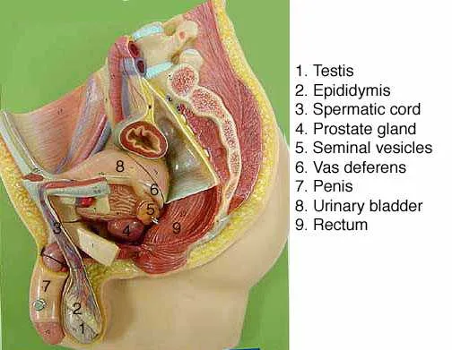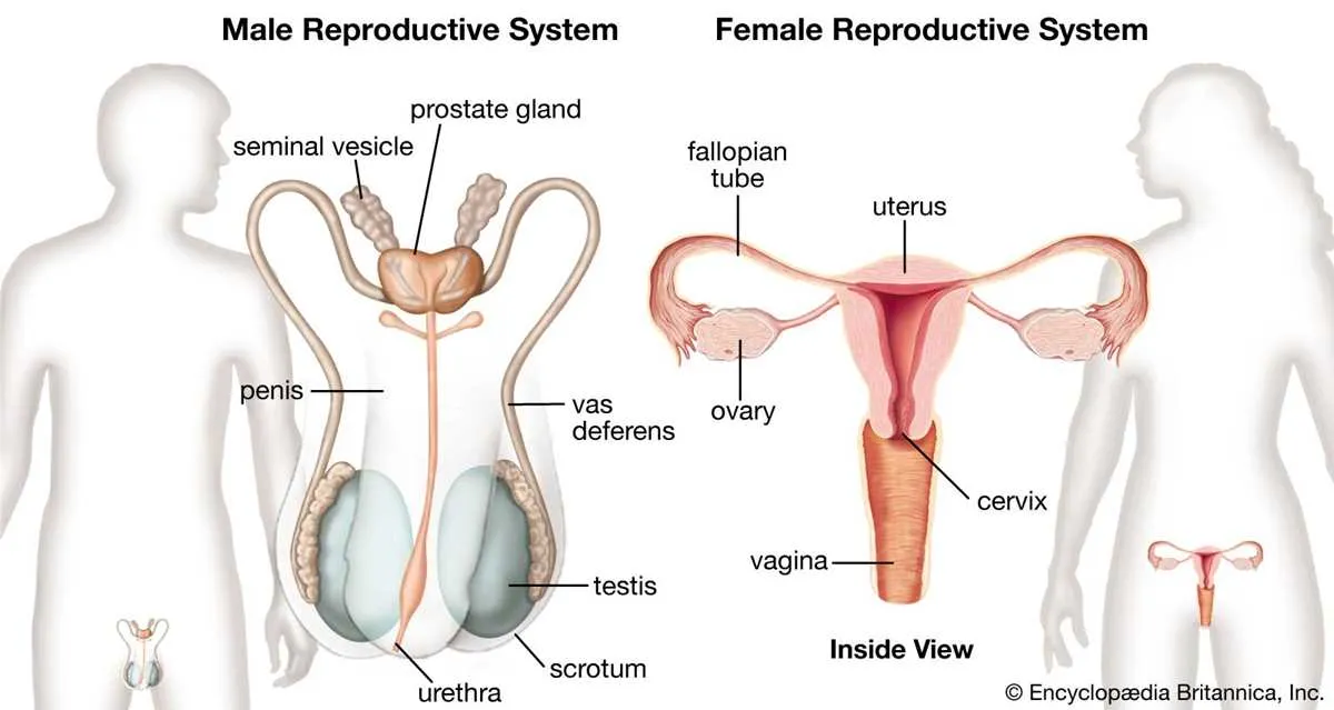
To gain a comprehensive understanding of the male genital organs, it’s crucial to familiarize yourself with their structure and functions. An effective way to do this is by studying a well-detailed illustration of the organs, highlighting each part’s unique role in the process of reproduction.
Focus on key structures such as the testicles, which produce sperm and testosterone, and the penis, essential for the delivery of sperm during intercourse. Pay attention to the epididymis, where sperm matures, and the vas deferens, which transports sperm from the testicles to the urethra. Additionally, the prostate gland and seminal vesicles are important for producing fluids that nourish and protect sperm.
Once you familiarize yourself with these components, you will better understand how they work together in the male fertility process. By analyzing diagrams that display these features clearly, it’s easier to visualize their interconnection and gain deeper insights into male sexual health.
Detailed Overview of the Human Anatomy: Reproductive Organs

Understanding the key components of the male anatomical structure is crucial for grasping how reproduction works. The essential organs are structured to facilitate the production and transport of sperm, along with other vital functions. Below is an overview of these components, with their respective labels for clarity.
| Organ | Function |
|---|---|
| Testes | Responsible for the production of sperm and testosterone. |
| Epididymis | A storage and maturation site for sperm cells. |
| Vas deferens | Transports sperm from the epididymis to the urethra. |
| Seminal vesicles | Produce a fluid that nourishes sperm and makes up a part of semen. |
| Prostate gland | Secretes fluid that helps sperm survive in the female reproductive tract. |
| Urethra | Serves as the passage for both urine and semen. |
| Penis | Facilitates the delivery of semen into the female reproductive tract during intercourse. |
Understanding the Structure of Male Reproductive Organs

The testicles are responsible for the production of sperm and testosterone, which are critical for fertility and sexual function. They are located in the scrotum, a pouch of skin that helps regulate temperature. The optimal temperature for sperm production is slightly lower than the body’s core temperature, which is why the scrotum adjusts its position to maintain the right environment.
The epididymis, situated behind the testicles, stores and matures sperm until they are ready for ejaculation. This structure is crucial for sperm development, as it allows them to gain motility and the ability to fertilize an egg.
The vas deferens is a tube that transports sperm from the epididymis to the urethra. During ejaculation, sperm mix with fluids from the prostate gland and seminal vesicles to form semen, which is then expelled through the penis. Proper function of the vas deferens is essential for successful fertilization.
The prostate gland produces a significant portion of the fluid that makes up semen. It also contains enzymes that protect sperm and help with their motility. Any issues with the prostate, such as enlargement or inflammation, can impact fertility and sexual health.
The penis is the organ through which semen is delivered during intercourse. It consists of three main parts: the root, shaft, and glans. The shaft contains erectile tissue that fills with blood during arousal, enabling the organ to become erect. The glans, or head, is the sensitive tip of the penis, which contains numerous nerve endings for sexual pleasure.
The urethra serves a dual purpose, carrying urine from the bladder and semen from the reproductive organs. It runs through the penis and is essential for both urination and ejaculation. Proper function of the urethra is necessary for both urinary and sexual health.
Functions of Each Component in Male Reproduction
The testes are responsible for sperm production and the secretion of testosterone, which regulates secondary sex characteristics and sperm maturation.
The epididymis stores and matures sperm cells, allowing them to gain motility before ejaculation.
The vas deferens transports sperm from the epididymis to the urethra during ejaculation.
The seminal vesicles produce seminal fluid, which provides nutrients and energy for sperm, aiding their mobility and longevity.
The prostate gland secretes a fluid that nourishes and protects sperm, ensuring optimal conditions for fertilization.
The bulbourethral glands release a pre-ejaculate fluid that neutralizes acidity in the urethra, creating a safer environment for sperm during passage.
The urethra serves as a passage for both urine and semen, with a valve mechanism that prevents both from mixing during ejaculation.
The penis delivers semen into the female reproductive tract during intercourse, facilitating fertilization.
How to Identify Key Features on a Male Anatomy Illustration

To accurately identify the primary components on an illustration of the male anatomy, focus on the following points:
- Testicles – Located in the scrotum, these are the oval-shaped glands responsible for sperm and testosterone production. They are positioned at the bottom of the image.
- Epididymis – A coiled structure at the back of each testicle. This is where sperm matures and is stored.
- Vas deferens – A long tube that transports sperm from the epididymis to the urethra. It is typically shown as a continuation from the epididymis leading towards the pelvic cavity.
- Seminal vesicles – Located near the base of the bladder, these glands contribute fluid to semen, providing nutrients and aiding sperm motility.
- Prostate gland – Positioned beneath the bladder, it surrounds the urethra and produces a fluid that forms part of semen. It is often shown as a small, round structure near the base of the bladder.
- Urethra – The tube that runs through the penis, facilitating the passage of urine and semen. It is typically depicted running through the central part of the diagram, with branches leading from the prostate.
- Penis – The external organ responsible for urination and sexual function, often depicted as a long, cylindrical shape extending from the body.
- Bulbourethral glands – Small glands that secrete a clear fluid into the urethra to neutralize any acidity, located near the base of the penis.
Use these landmarks to quickly orient yourself within the diagram, ensuring each part is correctly identified. Pay attention to the relative size and positioning of each structure, as this will help in distinguishing between similar organs.