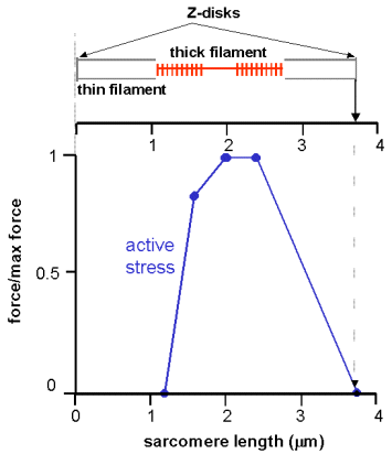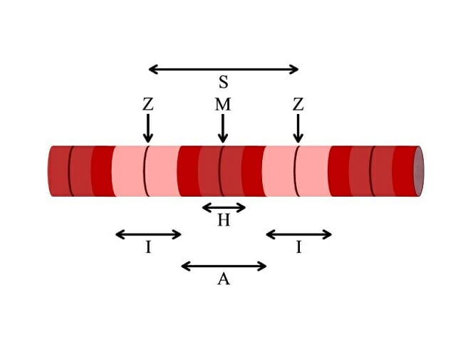
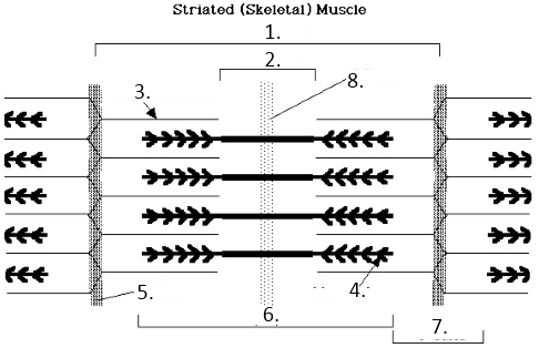
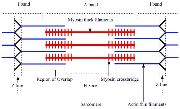
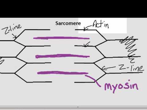
Sarcomeres are composed of thick filaments and thin filaments.
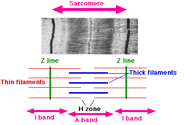
The thin filaments Look at the diagram above and realize what happens as a muscle contracts. Draw your own diagram of two sarcomeres.
The first should be of a relaxed muscle. The second should be of a contracted muscle.
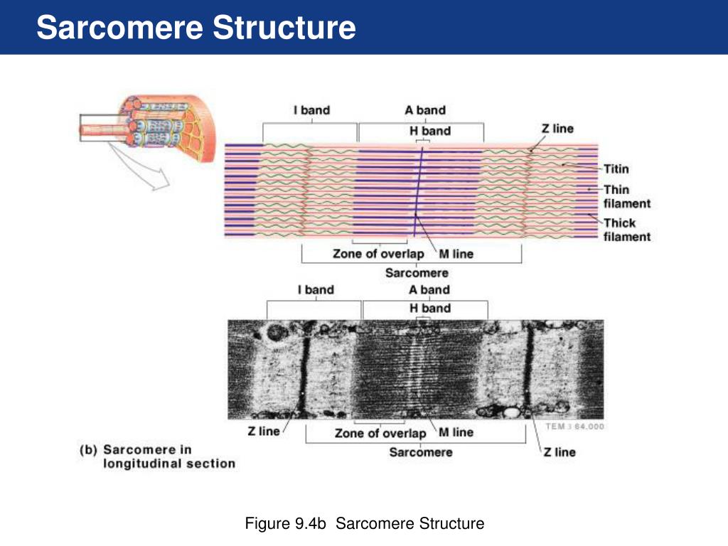
Label the Z line, M line. Start studying UNIT 5: Label the parts of the Sarcomere. Learn vocabulary, terms, and more with flashcards, games, and other study tools.
A sarcomere is the basic unit of striated muscle tissue. It is the repeating unit between two Z lines. Skeletal muscles are composed of tubular muscle cells which.
Each myofibril is made up of contractile sarcomeres AND Drawing labelled diagrams of the structure of a sarcomere.Summary. A little over 50 years ago, Sydney Brenner had the foresight to develop the nematode (round worm) Caenorhabditis elegans as a genetic model for understanding questions of developmental biology and neurobiology.
Over time, research on C. elegans has expanded to explore a wealth of diverse areas in modern biology including studies of the basic functions and interactions of eukaryotic.
Port Manteaux churns out silly new words when you feed it an idea or two. Enter a word (or two) above and you’ll get back a bunch of portmanteaux created by jamming together words that are conceptually related to your inputs..
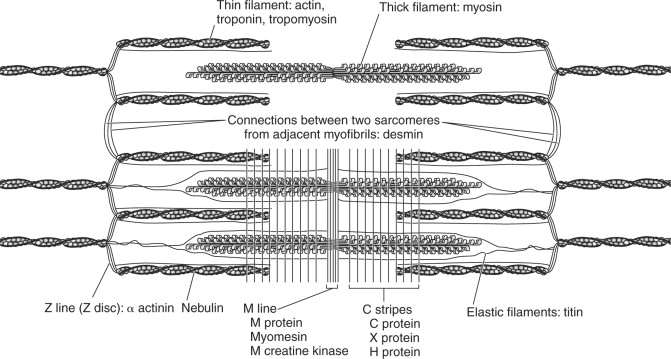
For example, enter “giraffe” and you’ll get . A myofibril (also known as a muscle fibril) is a basic rod-like unit of a muscle cell.
Muscles are composed of tubular cells called myocytes, known as muscle fibers in striated muscle, and these cells in turn contain many chains of schematron.org are created during embryonic development in a process known as myogenesis.. Myofibrils are composed of long proteins including actin, myosin, and.
Find muscle anatomy Stock Images in HD and millions of other royalty-free stock photos, illustrations, and vectors in the Shutterstock collection. Thousands of new, high-quality pictures added every day.
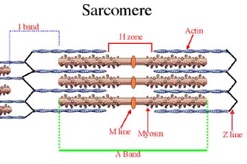
Fig 2: The C. elegans wiring diagram is a network of identifiable, labeled neurons connected by chemical and electrical synapses.
Red, sensory neurons; blue, interneurons; green, motorneurons. Signal flow view shows neurons arranged so that the direction of signal flow is mostly downward.Sarcomere – WikipediaMuscle Anatomy Images, Stock Photos & Vectors | Shutterstock
